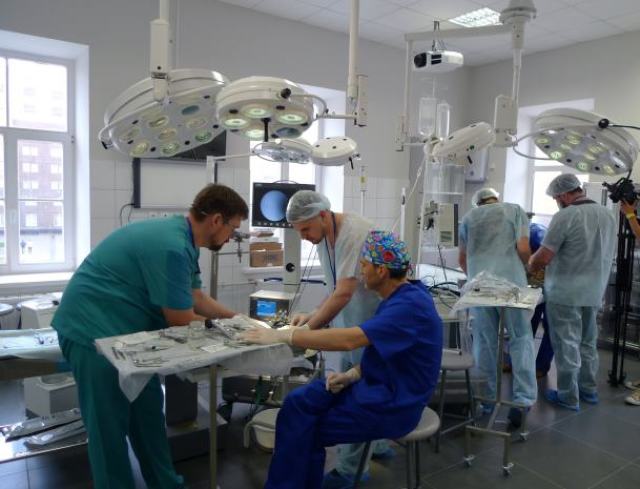
In the clinic of military traumatology and Orthopedics of the Military Medical Academy, more than a year ago, the 3D modeling method was successfully implemented and is actively used. It is designed to simplify the surgical operation, correct preoperative planning and practice the surgeon's actions on three-dimensional models.
This approach allows the doctor to examine the patient's fracture from all sides, as well as to outline the course of surgical actions. The specialists of the clinic, based on the data of computed tomography, make a three-dimensional sample that completely simulates a complex fracture. This allows surgeons to assess the severity of the injury and accurately measure all injuries.
In May 2021, the clinic of military Traumatology and Orthopedics successfully performed surgery on a patient with a closed intra-compound comminuted fracture of the distal metaepiphysis of the left humerus with displacement of fragments. The case was complicated by the multi-splintered nature of the fracture.
After the preoperative planning, the specialists of the clinic created a three-dimensional model of a complex fracture, which allowed them to visualize and work out the course of the operation in advance. The fracture model is an exact copy of the injury with the direction of the bone fragments and the degree of their displacement. Thus, after using 3D modeling, it turned out that only two fragments needed repassion, i.e. more surgical intervention. Thanks to this model, doctors were able to work out the necessary surgical interventions before starting the operation itself.
It is worth noting that in comparison with the traditional method, planning and working out complex operations using 3D-printed models reduces the risk of an unsuccessful operation and allows you to choose the best option for surgical intervention.
In addition, 3D modeling and samples with complex fractures are used in teaching the profession. The future doctor can not only study the nature of the damage in detail, but also apply practical skills, simulating the course of the operation without harming the patient.
The Ministry of Defense noted that before the advent of modern 3D modeling technologies, the academy's specialists made a forecast based on data obtained by computed tomography. The image from the images from the tomograph is presented in a flat format, which significantly reduces the quality of the development of the preoperative plan, and this, in turn, increases the time of the operation and increases the risks, including the development of postoperative complications.

